Treated by Dr. Ravi Chandra Reddy Obili at Dr Ravi Chandra Reddy
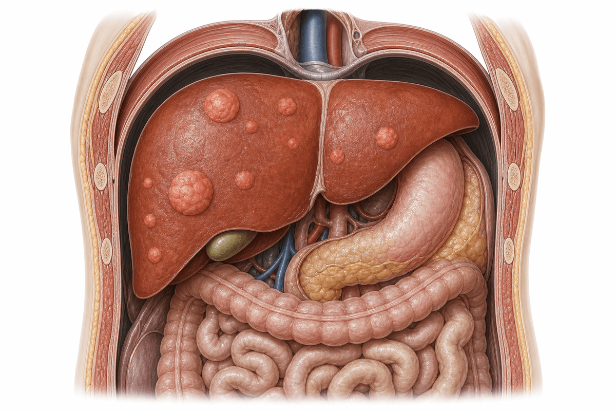
Benign liver tumours are non-cancerous growths in the liver that are commonly detected in Visakhapatnam through imaging studies. These tumours include hemangiomas, focal nodular hyperplasia (FNH), and hepatic adenomas, which usually grow slowly and rarely cause symptoms. Most cases can be managed conservatively, though some require surgical intervention by Dr Ravi Chandra Reddy Obili, an experienced surgical gastroenterologist who specializes in hepatobiliary conditions.
The most common benign liver tumour, composed of blood vessels. Typically asymptomatic and discovered incidentally during imaging. Usually requires no treatment unless large or symptomatic.
A benign nodular lesion caused by localized hyperplastic response to vascular malformation. Second most common benign liver tumour, more frequent in women. Generally stable and does not require intervention.
A rare benign tumour associated with oral contraceptive use, anabolic steroids, or metabolic conditions. Has potential for rupture, bleeding, or malignant transformation, often requiring surgical management.
Multiple factors can contribute to the development and progression of this condition.
Benign Liver Tumours develops gradually. Recognising symptoms early gives you more treatment options.
From conservative to surgical — we always start with the least invasive option first.
A structured, patient-first approach from first visit to full recovery.
Dr Ravi Chandra Reddy Obili conducts thorough clinical assessment including detailed history, physical examination, and review of imaging studies. Advanced contrast-enhanced MRI or triphasic CT scans are performed to characterize the tumour and differentiate between various benign liver lesions, ensuring accurate diagnosis before treatment planning.
Based on tumour type, size, location, and patient symptoms, Dr Obili develops individualized management plans. Small asymptomatic hemangiomas and FNH are placed on surveillance protocols, while adenomas are evaluated for risk factors requiring intervention. Each case is discussed with consideration of patient preferences and lifestyle factors.
For tumours requiring surgical management, Dr Ravi Chandra Reddy Obili utilizes advanced laparoscopic techniques whenever feasible to minimize invasiveness. Preoperative planning includes volumetric assessment, vascular mapping, and anesthetic optimization. Open surgery is performed when tumour complexity or size necessitates traditional approach for patient safety.
All patients receive structured follow-up care with scheduled imaging surveillance and clinical assessments. Post-surgical patients undergo recovery monitoring, wound care, and functional assessment. Those on active surveillance have protocol-based imaging at defined intervals to detect any changes requiring intervention, ensuring long-term optimal outcomes.
What to expect at each phase of recovery.
For surgical resection, patients typically stay in hospital for 3-7 days depending on extent of surgery. Pain management, early mobilization, and monitoring for complications are priorities. Laparoscopic procedures allow faster recovery with discharge often within 2-3 days. Drain management and wound care instructions are provided.
During the first 2-4 weeks post-surgery, patients gradually resume normal activities with restrictions on heavy lifting and strenuous exercise. Follow-up visits include wound assessment, liver function testing, and imaging to confirm surgical success. Most patients return to light work within 2-3 weeks after laparoscopic surgery.
Complete recovery from liver resection typically occurs within 6-8 weeks. Periodic imaging surveillance continues for at least 1-2 years to monitor for recurrence or residual disease. Liver function normalizes as the remaining liver regenerates. Patients maintain regular follow-up appointments with lifestyle counseling and preventive care guidance.
Benign liver tumours have excellent long-term outcomes with appropriate management. Hemangiomas and FNH rarely cause complications and most remain stable throughout life. Surgical resection when indicated provides definitive cure with low recurrence rates.
Surgical removal of symptomatic or high-risk benign liver tumours achieves complete resolution in over 95% of cases. Dr Obili's expertise in hepatobiliary surgery ensures optimal outcomes with minimal complications. Most patients experience significant symptom relief and improved quality of life post-operatively.
With proper surveillance and timely intervention for adenomas, the risk of malignant transformation is significantly reduced. Hemangiomas and FNH have virtually no malignancy potential. Regular monitoring protocols ensure any concerning changes are detected early.
Laparoscopic liver resection techniques result in shorter hospital stays, less post-operative pain, and faster return to normal activities. Most patients resume full activities within 4-6 weeks. Cosmetic outcomes are superior with minimal scarring and excellent patient satisfaction.
Untreated benign liver tumours carry specific risks depending on type. Hepatic adenomas larger than 5 cm have risk of spontaneous rupture causing life-threatening intraperitoneal hemorrhage, and rare potential for malignant transformation to hepatocellular carcinoma. Large symptomatic tumours can cause progressive pain, compression of adjacent organs, and significantly impaired quality of life requiring emergency intervention.
Seek immediate medical attention if you experience sudden severe abdominal pain, signs of internal bleeding such as dizziness or rapid heartbeat, or a palpable abdominal mass. Schedule consultation with Dr Ravi Chandra Reddy Obili if you have been diagnosed with a liver lesion on imaging, have persistent right upper quadrant discomfort, or have risk factors such as oral contraceptive use with known liver adenoma. Early evaluation allows appropriate risk stratification and timely intervention when indicated.
Early treatment means more options and better outcomes. Book a consultation to understand your condition and explore the right path forward.